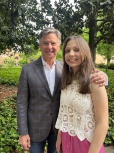
Dr. Dan Barrow & Elle
Our foremost objective is to advance research aimed at finding a cure for Elleanor’s rare disease. Simultaneously, there are medications and technologies currently undergoing research and development here in Atlanta that offer the potential to not only aid Elle, but also to provide widespread benefit to those suffering from a variety of brain disorders.
The partnership between Emory University’s surgeon-scientists and Georgia Tech’s biomedical engineers promises to expedite research, paving the way for groundbreaking solutions to numerous neurological disorders impacting humanity
This new collaboration is focused on improving brain function for a wide variety of disorders, such as ALS, Parkinson’s, Alzheimer’s, traumatic brain injury and stroke. And the innovations in surgical technologies and techniques promise to make “inoperable” brain tumors a thing of the past.
One of the most important collaborations in neurosciences is the interaction that occurs among neurosurgeons at Emory University and the biomedical engineers at the Georgia Institute of Technology. Emory and Georgia Tech have a combined Department of Biomedical Engineering that is ranked among the top in the nation. Emory, the sole major academic medical center in the Atlanta metro area (population of nearly 7 million residents), boasts access to a vast patient pool. Meanwhile, Georgia Tech houses some of the globe’s brightest biomedical engineers. By collaborating, Emory’s surgeon-scientists and Georgia Tech’s biomedical engineers can significantly expedite research progress, pioneering innovative solutions for numerous neurological disorders impacting humanity.
Elle’s Angels Atlanta has pledged financial support to this team of top-tier medical collaborators, led by Emory University’s Department of Neurosurgery Chair, Dr. Dan Barrow
Medical research is often performed by scientists working in isolation. When doctors work in collaboration with scientists, their research can focus on specific challenges faced by their human patients. This collaboration markedly increases the likelihood of research being applicable to conditions affecting humankind. Additionally, surgeons have the unique opportunity to not only access the human brain during surgical procedures, but to obtain tissue removed during brain tumor resections, epilepsy surgery, and other procedures. This unique perspective and these specimens sampled during surgery, can be used to better understand the function and dysfunction of the human brain.
Of particular interest to Elle’s Angels is the work this team is doing to restore function following stroke and traumatic brain injury. These functional deficits closely resemble the effects experienced by patients with Elle’s cerebrovascular disease (CCM).
Using an array of micro-electrodes, as well as brain-to-computer interfaces, the team has been working to restore limb functions, along with restoring signals from the motor cortex which have been disconnected during trauma, in cases such as traumatic brain injury and stroke. Brain and spinal cord injuries destroy motor neurons, and thus, a person’s ability to function. Emory’s investigational system is allowing patients to control a computer cursor simply by thinking about the movement of their own paralyzed limb, directly driving limb movement by stimulating electrodes.
This past December (2023) the Emory/Georgia Tech team lead by Nick Au Yong, MD, PhD, (Emory Functional & Restorative Neurosurgeon and Biomedical Engineering), and Chethan Pandarinath PhD (Biomedical Engineering & Neurosurgery) successfully implanted a brain-computer interface (BCI) into their first patient, a woman with a brain stem stroke who is paralyzed and has slurred speech. She was implanted with recording arrays in an area of the brain that controls her hand movement and speech areas. This surgery represents the culmination of years of work, including the recruitment of Dr. Pandarinath (a neural engineer recruited from Stanford University) to build the first multidisciplinary BCI program in Georgia.
Micro Robotics to Treat Brain Tumors (Dr. Kimberly Hoang):
Malignant brain tumor patients still have a very poor, terminal prognosis (approximately 1.5 years from time of diagnosis to death) despite all the advances we have made. The technological tools at a neurosurgeon’s disposal are limited, making craniotomies (removing part of the skull to expose the brain for surgery) and tumor resection dangerous. And then there are tumors too deep, or in too sensitive an area (brain stem, spinal cord) to safely remove, without damaging surrounding areas responsible for critical function such as movement, vision and breathing. Dr. Hoang’s lab is developing minimally invasive nanoscale robots (smaller than 1mm in size) that have the potential to perform local microsurgeries targeting areas of the brain currently out of the surgeons reach. These magnetically actuated microbots easily travel over the uneven surface of the brain, and within fluid-filled ventricles in controlled, non-linear trajectories. Current robot technology often uses macroscale instruments that are limited to linear trajectories to access brain tumors, and their use requires craniotomies, making surgery risky and highly invasive. Thus, the untethered steering device being created at Emory has the potential to reach regions of the brain currently inaccessible.
This technology also has tremendous potential for transporting drug delivery and gene therapy for most any neurological, psychiatric or neurodegenerative disorder imaginable. It’s hard to deliver drugs to the brain due to both the blood-brain barrier and the risks of repeated surgeries and interventions. Dr. Hoang’s lab has shown that these microbots can deliver tiny packets of drugs for targeted drug delivery. By carrying “backpacks” made of smart hydrogels, when the acidity of the surrounding area changes, as is the case with brain tumors, the backpacks swell. This swelling releases nanoparticles in the backpack loaded with the desired drug. These microbots equipped with backpacks can be controlled individually or operate collectively in a “swarm, executing intricate tasks seamlessly.
Opening the Blood-Brain Barrier:
Neurological disorders account for more than 6% of the global disease burden, with costs in the US alone exceeding $1 trillion per year. The blood-brain barrier is like an iron door, preventing 98% of all potential pharmaceuticals from reaching the central nervous system. Emory is interested in investing in a low-frequency transducer ($1M) which would interface with their high-intensity focused ultrasound machine to selectively open the blood-brain barrier. This would allow researchers to explore the possibility of breaking up Alzheimer’s plaques, shrinking brain tumors, creating more effective low-dose pharmacological options, and experimenting with gene therapies for a host of neurological and neurodegenerative conditions.
Therapeutic Strategies for Traumatic Brain Injury (Dr. David Gimbel):
Currently, there is no effective therapeutic treatment for brain injury. Emory researchers studying traumatic brain injury are working with Grady Memorial Hospital’s Neurotrauma center to identify the striking similarities of the cellular prion protein common in both traumatic brain injury and Alzheimer’s disease. This discovery of the similarities could use Alzheimer’s therapies to inform a preventative therapeutic means of protecting contact-sport athletes. It might also be possible to develop a neuroprotective drug that can be given to TBI patients immediately after incurring injury…making it the first-ever preventative drug for brain injury. Dr. Gimbel runs a neurotrauma lab at Emory where he is using existing Alzheimer’ therapeutics in mouse models for brain injury research. These treatments seem to completely protect mice from injury, as the therapeutically-treated injured mice appear to have not had any injury at all. These molecules, initially created for Alzheimer’s disease, may become the first ever effective therapeutic for head injury victims.
Targeting Stem Cells to Prevent Tumor Recurrence (Dr. Tomas Garzon-Muvdi):
Glioblastoma is a very aggressive tumor, resistant to chemotherapy and radiation. Additionally, glioblastoma cells can migrate to the healthy brain, as well as return following an otherwise successful resection. And they are notoriously resistant to current therapies. Emory is working on ways to decrease glioblastoma cell invasion into the healthy brain, as well as finding ways to target the stem cell population in an effort to prevent origination, recurrence and therapy resistance. Emory’s novel research indicates that a particular protein implicated in other organ cancers has decreased expression in brain tumor stem cells. By targeting this protein and overexpressing it, researchers hope to kill those deadly stem cells.
Microsensors to Distinguish Healthy Brain Tissue from Tumors (Dr. Kimberly Hoang):
Distinguishing between healthy brain tissue and abnormal lesions is challenging for even the most experienced neurosurgeons. They currently must rely on contrast dye to visualize brain matter, which is not only subjective, but complicated by tumors that are dye resistant. Furthermore, because brain tissue is responsible for so many delicate functions, it is difficult to take margins when extracting a tumor and difficult to know in real-time exactly what to remove and what to remain intact. Emory’s team is in the process of developing microsensors to quantitatively distinguish healthy tissue from tumors, making neurosurgery safer and more effective. In partnership with Georgia Tech, Emory’s research team is working to develop a flexible arm that can replace the linear tools that are so limiting.
Alterative Treatment Options for Diffuse Intrinsic Pontine Gliomas (Dr. Kimberly Hoang):
Approximately 20% of all pediatric brain tumors are Diffuse Intrinsic Pontine Gliomas (DIPG), a deadly form of pediatric brain cancer that occurs in the pons, the part of the brainstem that links the brain to the spinal cord. These tumors (Elleanor’s original diagnosis) usually develop in children when they are 5-7 years old. There are no viable treatment options, and due to the highly sensitive nature of the brainstem, most are never biopsied since severe neurological complications are likely. As these tumors are typically inoperable, there is a need for alternative treatment approaches, including direct delivery of chemotherapeutic agents into the masses. Effective therapy research for these aggressive brain tumors is crippled by the absence of a good large animal model in which surgical approaches can be refined. Emory has developed the first pig model to support research for DIPG interventional approaches. There is now an opportunity to develop a more advanced method of neurosurgical navigation, and Philanthropy would enable Emory to evaluate the efficacy of new proposed therapeutic strategies.